

2024
Apr
24
Welcome to Immunology
The purpose of the Immunology Program is to provide education leading to a Ph.D. in Immunology. This Program is under the responsibility of the Committee on Immunology at Harvard. The Committee includes over 130 faculty representing a broad area of research interests including transplantation, neuro-immunology, autoimmunity, stem cell biology, infection and immunity, human translational immunology, tumor immunology, immunobiology and mucosal immunity.
Our goal is to educate scientists in investigative and academic medicine, preparing them to contribute to immunological research with a full awareness of the potential impact of immunology. Our program combines an education in basic biology, a sophisticated training in immunology, and exposure to the immunological and non-immunological problems of disease.
PhD Graduate Alexandra Schnell Receives 2023 Weintraub Award

Our very own IMM recent PhD Graduate, Dr. Alexandra Schnell,
was one of the recipients of the 2023 Harold M. Weintraub Award.
Click here to view the complete article.
Wednesday Seminar Series
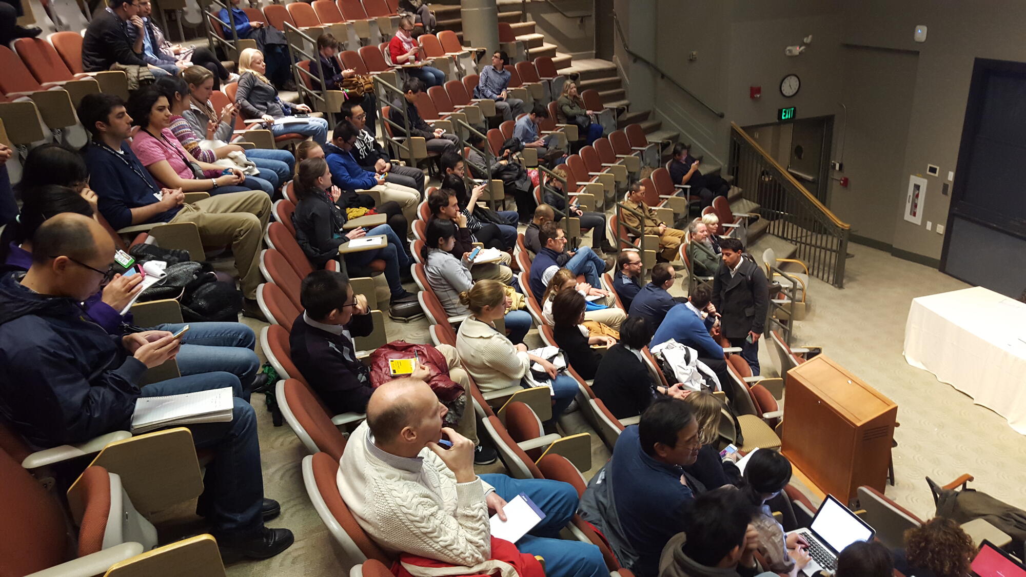 The Wednesday Seminar Series is one of the most widely-attended in the Longwood Medical area and features presentations by leaders in the field of Immunology. View a complete schedule.
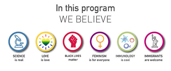
Figure Inspired by the Diversity and Inclusion Lab Posters of Sammy Katta